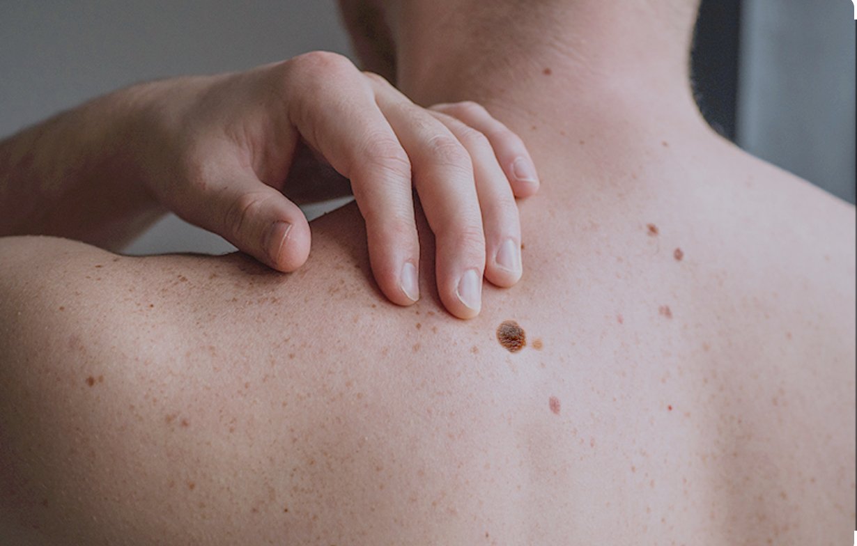
MELANOMA
Types, causes, symptoms, stages and treatment.
Melanoma is a type of skin cancer which although relatively uncommon as compared to other types of skin cancers, is more dangerous as its more likely to spread. Its a malignant tumour of melanocytes (pigment-producing skin cells) that usually arises on the skin but can also occur in the eye or mucous membrane.
TYPES OF MELANOMA
- Superficial spreading melanoma (≈70%): The most common type; often arises in an existing mole and spreads outward in the top layer of skin
- Nodular melanoma (≈20%): Grows as a raised bump (nodule) and often invades more deeply; tends to be more aggressive
- Lentigo maligna melanoma: Develops from a lentigo maligna (a long-standing sun-damaged patch) in older adults, grows slowly at first
Acral lentiginous melanoma: Appears on non–sun-exposed areas (palms, soles, under nails); more common in darker-skinned individuals
MAJOR CAUSES OF MELANOMA
- Ultraviolet(UV) Exposure: Intense intermittent sun exposure, blistering sunburns, and use of tanning beds greatly increase melanoma risk . UV radiation induces DNA mutations in skin cells, notably in the BRAF oncogene (mutated in ~50% of melanomas) .
- Pigmentation and Skin Type: Fair skin, light hair (blond/red), and light eye color are associated with higher risk. These traits confer less natural protection against UV damage.
- Nevus (Mole) Count and Type: Having many moles, or atypical/dysplastic nevi, raises risk. Melanoma often arises de novo or in a pre-existing mole. The “ugly duckling” sign (a mole that looks markedly different from one’s others) is a warning sign.
- Family History / Genetics: A family history of melanoma (or pancreatic cancer) indicates possible inherited risk. High-risk familial melanoma often involves germline mutations in tumor-suppressor genes such as CDKN2A (p16), CDK4 or BAP1 . These inherited mutations markedly increase melanoma risk (lifetime risk ~28–67% for CDKN2A carriers) . Rare conditions like xeroderma pigmentosum (DNA repair defect) also confer very high risk .
- Personal History: A prior melanoma or non-melanoma skin cancer increases recurrence risk. Immunosuppression (HIV infection, organ transplant, chronic immunosuppressive drugs) also elevates melanoma risk .
Age and Gender: Although melanoma can occur at any age, risk rises with age. In older adults, risk is higher in men; in younger adults, especially women, cases are also common for unclear reasons .
SIGNS AND SYMPTOMS
- New or changing pigmented lesion: Any new spot or mole, or a mole that enlarges, changes shape, color, or elevation. The ABCDE rule can help spot suspicious lesions (Asymmetry, Borders, Colour, Diameter, Evolving/elevation)
- “Ugly duckling” sign: A mole that looks noticeably different from the patient’s other moles .
- Other skin changes: A sore that doesn’t heal, spread of pigment from the border of a spot into surrounding skin, or new swelling, tenderness or itching of a mole. Any mole that bleeds or becomes painful warrants evaluation.
Location-specific signs: Melanoma under the nail (pigmented streak or ulceration of a nail bed) or on scalp, genitalia, or mucosa may be less obvious but follow the same ABCDE changes.
DIAGNOSTIC PROCEDURES
Melanoma diagnosis usually relies on histologic examination of the suspicious lesion with imaging if its spread. Skin biopsy, Sentinel lymph node biopsy (SNLB), Imaging studies, molecular testing and dermatology are the major steps involved in diagnosis of melanoma to establish the pathological stage and guide treatment.
STAGES IN MELANOMA
- Stage 0 (melanoma in situ): Confined to the epidermis (Tis, N0, M0) .
- Stage I (localized): Tumor ≤2.0 mm thick (with or without ulceration) and no nodal spread (e.g. T1 or T2a, N0, M0). (Stage I is subdivided into IA and IB by thickness and ulceration.)
- Stage II: Tumor >2.0 mm thick (up to any depth, possibly ulcerated) and no nodal spread . Stage II is further divided (IIA, IIB, IIC) by thickness (up to 4 mm, >4 mm) and ulceration status .
- Stage III: Any thickness tumor with regional nodal involvement or in-transit metastases, but no distant metastasis . Subcategories IIIA-IIID depend on number of nodes and ulceration.
- Stage IV: Melanoma has spread to distant sites (distant skin, lymph nodes, or visceral organs)
TREATMENT
- Surgery: Mohs micrographic surgery is occasionally used for very thin melanomas in cosmetically sensitive areas . If SLNB is positive, completion lymph node dissection may be offered, although recent practice may favor observation or adjuvant therapy instead. Surgery may also remove isolated metastases for palliation or potential benefit.
- Immunotherapy: Checkpoint inhibitors have revolutionized melanoma treatment. PD-1 inhibitors (pembrolizumab, nivolumab) and CTLA-4 inhibitor (ipilimumab) unleash the immune system against melanoma. These drugs are used for unresectable or metastatic melanoma, and increasingly as adjuvant therapy after surgery for high-risk Stage II/III
- Targeted Therapy: For melanomas with specific mutations, targeted drugs can be highly effective. About 50% of cutaneous melanomas harbour a BRAF^V600 mutation . BRAF inhibitors and MEK inhibitors are used in combination for metastatic BRAF-mutant melanoma.
- Chemotherapy: Traditional cytotoxic drugs are now rarely first-line, given the success of immunotherapy/targeted therapy. Agents include dacarbazine, temozolomide, platinum compounds, and taxanes . These may be used if other therapies fail or are contraindicated. Regional chemotherapy (isolated limb perfusion/infusion with high-dose melphalan) can treat in-transit limb metastases
- Radiation Therapy: Melanoma is relatively radioresistant, so radiation is not routinely used for primary lesions. However, radiation can be valuable in certain situations :
- Adjuvant treatment of high-risk areas (e.g. after resection of many positive nodes, or desmoplastic melanoma which tends to recur locally).
- Palliation of metastatic disease (brain, bone, or other sites) to relieve symptoms .
Local control of satellite or in-transit metastases when surgery is not feasible
PROGNOSIS AND SURVIVAL RATES
- Prognosis depends strongly on stage at diagnosis. Overall, melanoma has high survival if caught early, but drops sharply with metastasis. Five-year relative survival (based on US SEER data) is approximately :
- Localized melanoma (confined to skin, roughly Stage 0/I): >99% survive 5 years .
- Regional melanoma (spread to nearby lymph nodes or structures, Stage III): ~75% 5-year survival .
- Distant metastatic melanoma (Stage IV): ~35% 5-year survival
PREVENTION AND EARLY DETECTION
Melanoma risk can be greatly reduced with sun-safe behaviors, and early detection is critical for cure. Some recommendations include:
Sun protection: Limit UV exposure by seeking shade, wearing sun-protective clothing (long sleeves, wide-brimmed hat), and applying broad-spectrum sunscreen (SPF ≥30) liberally
Avoid tanning beds: Artificial UV from tanning beds/sunlamps is proven to increase melanoma risk, especially if used before age 30
Skin self-exam: Learn your skin and perform a full-body self-exam monthly . Look for new moles or changes in existing spots (using the ABCDE criteria). Report any suspicious lesion to a doctor immediately.
Professional skin exams: Have regular skin exams by a dermatologist or primary doctor, especially if you have high-risk features (personal/family history of melanoma, many atypical moles). Dermatoscopic examination and total-body photography can help track changes


